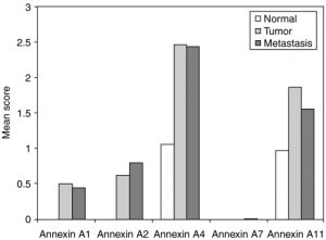
(Normal, colorectal tumor, and metastasizing colorectal cancer cells analyzed for intensity of annexin protein staining.)
1)The TITLE you would give these figures. Make it a single declarative statement about the data.
The cancer cells have higher annexin protein than the normal cells.
2) The background that the authors must have had in mind that made them ask this question. To do this, use the review article you read for last class.
MMPs break down ECM which pave the way for tumor cells to rapidly grow and enhancing the blood vessels (good environment for tumor growth) which also help the growth of tumor cells. Annexin helps to regulate MMPs, and therefore controls tumor growth.
3) The question asked. What were the researchers trying to figure out that led them to gather these data?
Which type of annexin(annexin A1, annexin A2, annexin A4, annexin A7, or annexin A11) have stronger protein staining on tumor and metastasis cells?
4) The methods employed. What were the subjects, the manipulations, the controls, the data collected?
Subjects: dead patients
Manipulations:
Independent variable: human cell
Dependent variable: intensity (color)
Control: normal human cells
Data collected: intensity of annexin protein staining in different type of cells (normal, colorectal tumor, and metastasizing colorectal cancer cells).
They extracted cells from dead bodies, applied stains on the cells, and observed the intensity stain of different types of cells (normal, colorectal tumor, and metastasizing colorectal cancer cells) under a microscope, to observe the intensity of annexin protein staining.
5) The observations made. What did they SEE
The highest and second highest percentage of the intensity of protein staining across the three cells are in Annexin A4 and A11 respectively. Within each Annexin A1, A4, and A11, tumor cells have the highest percentage of intensity staining. The percentage of intensity staining of normal cells in Annexin A4 and Annexin A11 are slightly similar. In Annexin A7, there is no visible intensity of protein staining in normal and collateral cells and barely any in metastasis cells.
6) The conclusions reached. Don’t OVER-reach – but how can one interpret these observations to address the question you imagine the researchers asked?
According to the graph, we can infer that there is more of Annexin A4 compared to the other types of Annexin in normal, colorectal tumor, and metastasizing colorectal cancer cells. There tends to be more Annexin in tumor and metastasizing colorectal cancer cells than normal cells, but this may not be the case in Annexin A7.
